
We’re thrilled to have our director of pathology, Sabrina McGraw, DVM, Ph.D., answer a few questions about the exciting new changes coming to AmplifyBio’s pathology lab. Here, she’ll discuss how cutting-edge technology is not only modernizing her lab but enabling better partnerships with clients as well.
Please describe your background in preclinical research and pathology. What led you to AmplifyBio?
Throughout my academic development, I had a focus in research in biology, pharmacology, and pathology. My Master’s degree focused on spontaneous models of neurologic disease in animal models, while my PhD centered on the pathology of infectious diseases. During my Anatomic Pathology residency, I worked in viral discovery, including in situ hybridization and other visualization techniques in preserved tissue sections. Following my board certification, I joined the U.S. Army, managing research programs and teams at the US Army Medical Research Institute of Chemical Defense (USAMRICD) and US Army Medical Research Institute of Infectious Disease (USAMRIID). After leaving the Army, I signed on to lead the team that would become AmplifyBio Pathology.

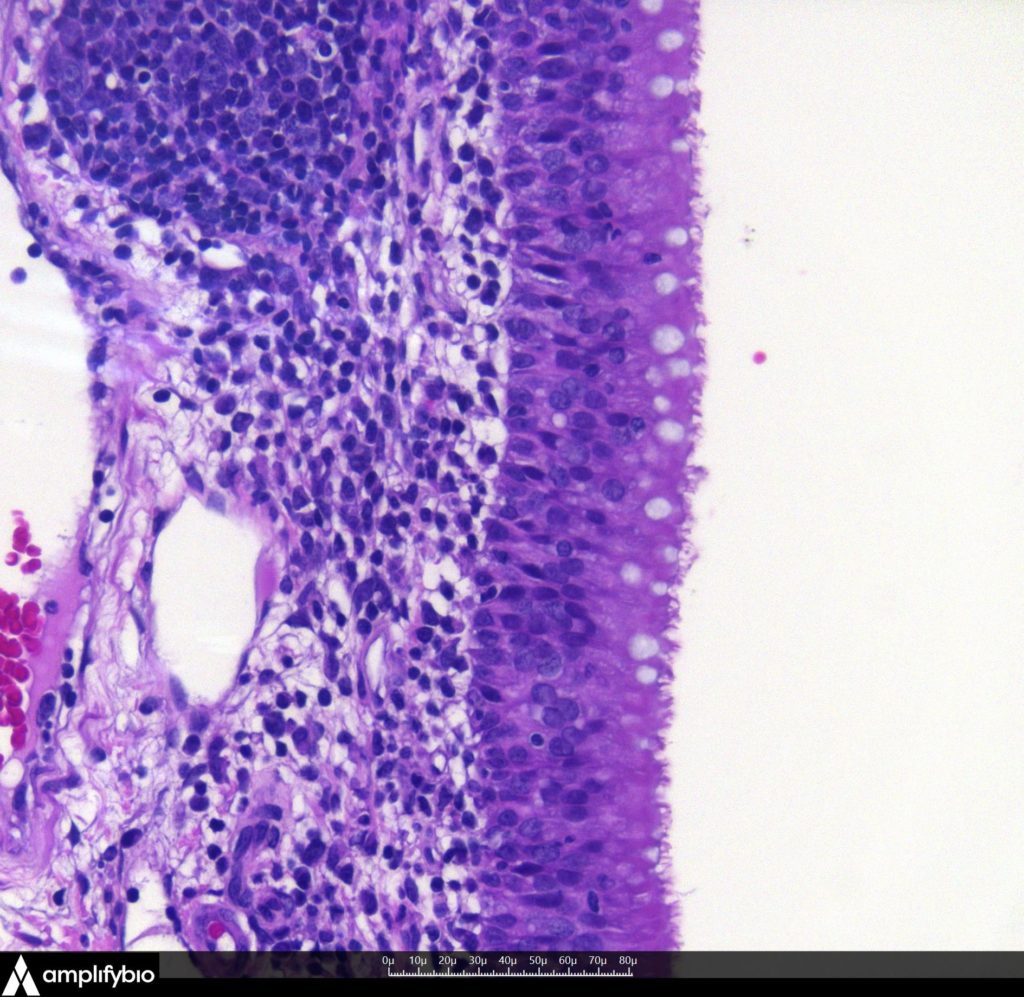
Immune stimulation of the nasopharyngeal mucosa. Note the ciliated respiratory epithelium overlying this tissue. Image captured using Augmentiqs camera and software.
What new technologies at AmplifyBio are you really excited about?
A picture may be worth a thousand words, but that is even more true in the world of Pathology! Our new (October 2022) Augmentiqs microscope camera systems enable our pathologists to take instant images of histopathology findings, which are then available to illustrate results, share with remote or regulatory pathologists, or affix to sponsor reports. This system reads a QR code label on the slide, embedding study and animal information, and even the objective magnification, directly into the image file metadata. Ultimately, this means every study will have well documented image files of pertinent or even background findings available in an instant.
In the coming months, we’ll have a new whole slide digitizer (Hamamatsu NanoZoomer) which creates whole slide image files with magnification levels of 2x to 60x and enables immunofluorescent labeling! This will allow for biodistribution evaluation as well as characterizing cell types and a myriad of other functionalities. Additionally, our histology group will be adding an immunohistochemistry stainer (Leica BOND Rx), which will provide high-throughput IHC capabilities plus in-situ hybridization (ISH), miRNA, TUNEL, bDNA, and multiplexing capabilities. In 2023 we’re bringing on an automated camera system in necropsy that will allow you to see necropsy findings in real time or allow the prosector to create training or study images or video using voice commands to keep their hands free.
How do these changes affect the clients’ studies and enable us to better collaborate on pathology?
Efficient, high quality image systems that provide reliable documentation of study and sample information enable the capture of images without impinging on study timelines. This elevates the interactions between pathologists, sponsors, and study teams, as well as providing a visual, searchable database of findings from a clients’ previous studies with a particular compound, which ensures consistency of results, with ready comparability. New capabilities in immunofluorescence, immunohistochemistry, and other marker visualization technologies will greatly improve our in-house ability to conduct the breadth of evaluations our clients need.
Are there any other changes being made to optimize your lab?
AmplifyBio is investing heavily in modernizing sample management, equipment monitoring, and digital file storage, and this is being especially felt in Pathology. Excellent documentation of equipment maintenance, validation, and calibration is a laborious process, and modernization efforts are already reducing employee time and reducing errors. Adding QR code labels, created in Provantis and including all pertinent study data, to tissue slides enable camera systems to document images and store them with embedded metadata. This even includes a snapshot of the slide itself, taken automatically while on the microscope, so that image data can be QC’d to the actual slide image.
What direction do you think all these modernization efforts will take us?
All these new imaging capabilities will be opening new doors for Amplifyio to move into digital image analysis, even including artificial intelligence. The first effort in this direction is Visiopharm, which we will be utilizing for stereologic evaluation of lung sections for emphysematous change. Our team has the talent and drive to become industry leaders in histopathologic evaluation. My goal is to have us focus on digitizing our current work, then bring in a greater breadth of techniques (IHC, ISH, IF, etc.). Then we move to digital analysis and beyond!
Read More:
Utilizing Analytics to Empower the Study Process
AmplifyBio CRO Pathology Capabilities